UV-induced fluorescence (UVIF) data that were obtained in the 1978 investigation of the Turin Shroud, were recently revisited by Pellicori supporting different behavior of background, non-image, and body image areas. Based on the experimental literature results, the chemical nature of that difference is discussed here. Former enzymatic analysis using pectinase indicated the presence of pectic substances, but only for fibers extracted from the background non-image areas. On the other hand, the high content of pectins in the primary cell wall (PCW) of flax fibers is well documented. The PCW is a thin outermost layer of fibers of approximately 0.2 μm, which corresponds to the layer where the body image is seen. Pectins are easier oxidized and destroyed than cellulose, the main component of the linen. In the present paper, pectins from the PCW of the linen fibers are proposed to participate in the initial stage of body image formation on the Turin Shroud, although they have been destroyed in that process. To demonstrate the presence of pectins in the fibers from the Shroud, the results of Fourier Transform Infrared (FTIR) spectroscopy in attenuated total reflectance (ATR) mode obtained by Fanti for body image and non-image fibers have been compared. Analyzed spectra do not exclude the presence of pectins. Thus, participation of pectins can explain some unique properties of the Shroud image, i.e., superficiality of the body image and nonuniform distribution of its yellow color.
| Published in | International Journal of Archaeology (Volume 13, Issue 2) |
| DOI | 10.11648/j.ija.20251302.15 |
| Page(s) | 178-184 |
| Creative Commons |
This is an Open Access article, distributed under the terms of the Creative Commons Attribution 4.0 International License (http://creativecommons.org/licenses/by/4.0/), which permits unrestricted use, distribution and reproduction in any medium or format, provided the original work is properly cited. |
| Copyright |
Copyright © The Author(s), 2025. Published by Science Publishing Group |
Fluorescence, FTIR Spectra, Turin Shroud, Pectins, Image Superficiality, Image Striations
ATR | Attenuated Total Reflectance |
CIE L | International Commission on Illumination Lab Color Space |
FTIR | Fourier Transform Infrared |
HG | Homogalacturonan |
PCW | Primary Cell Wall |
RGB | Red, Green, Blue Pixels |
STURP | Shroud of Turin Research Project |
UVIF | Ultraviolet Induced Fluorescence |
| [1] | Morris, R. A., Schwalbe, L. A., London, J. R. X-ray Fluorescence Investigation of the Shroud of Turin. X-Ray Spectrometry. 1980, 9(2), 40-47. |
| [2] | Pellicori, S. F. Spectral Properties of the Shroud of Turin. Applied Optics. 1980, 19(12), 1913-1920. |
| [3] | Gilbert, R. Jr., Gilbert, M. M. Ultraviolet-visible Reflectance and Fluorescence Spectra of the Shroud of Turin. Applied Optics. 1980, 19(12), 1930-1936. |
| [4] | Heller, J. H, Adler, A. D. Blood on the Shroud of Turin. Applied Optics. 1980, 19(16), 2742-2744. |
| [5] | Pellicori, S. F., Evans, M. S. The Shroud of Turin Through the Microscope. Archeology. 1981, 34, 34-43. |
| [6] | Miller, V. D., Pellicori, S. F. Ultraviolet Fluorescence Photography of the Shroud of Turin. Journal of Biological Photography. 1981, 49(3), 71-85. |
| [7] | Heller, J. H., Adler, A. D. A Chemical Investigation of the Shroud of Turin. Canadian Society of Forensic Science Journal. 1981, 14(3), 81-103. |
| [8] | Schwalbe, L. A., Rogers, R. N. Physics and Chemistry of the Shroud of Turin. A Summary of the 1978 Investigations. Analytica Chimica Acta. 1982, 135, 3-49. |
| [9] | Jumper, E. J., Adler, A. D., Jackson, J. P., Pellicori, S. F., Heller, J. H., Druzik, J. R. A Comprehensive Examination of the Various Stains and Images on the Shroud of Turin. Archeological Chemistry III, ACS Advances in Chemistry, Lambert, J. B., Ed., American Chemical Society, Washington D. C.; 1984, 205, 447-476. |
| [10] | Baima Bollone, P., Jorio, M., Massaro, A. L. La Dimostrazione della Presenza di Tracce di Sangue Umano sulla Sindone. Sindon. 1981, 30, 5-8. |
| [11] | Baima Bollone, P., Gaglio, A., Grillo, C., Zanin, A. Ricerca degli Antigeni M, N ed S Nelle Tracce di Sangue sulla Sindone. Sindon. 1985, 34, 9-13. |
| [12] | Fanti, G., Zagotto, G. Blood Reinforced by Pigments in the Reddish Stains of the Turin Shroud. Journal of Cultural Heritage. 2017, 25, 113-120. |
| [13] | Laude, J-P., Fanti, G. Raman and Energy Dispersive Spectroscopy (EDS) Analyses of a Micro Substance Adhering to a Fiber of the Turin Shroud. Applied Spectroscopy. 2017, 71(10), 2313-2324. |
| [14] | Di Lascio, A., Di Lazzaro, P., Iacomussi, P., Missori, M., Murra, D. Investigating the Color of the Blood Stains on Archeological Cloths: the Case of the Shroud of Turin. Applied Optics. 2018, 57, 6626-6631. |
| [15] | Fanti, G. New Insights on Blood Evidence from the Turin Shroud consistent with Jesus Christ’s Tortures. Archives of Hematology Case Reports and Reviews. 2024, 9(1), 1-15. |
| [16] | Pellicori, S. UV Fluorescence Imagery of the Turin Shroud – Digitally Revisited. International Journal of Archeology. 2020, 8(2), 32-36. |
| [17] | Schwalbe, L., Pellicori, S. Analysis of Photoelectric Colorimetry and Fluorimetry of the Turin Shroud. International Journal of Archeology. 2023, 11(1), 1-8. |
| [18] | Pellicori, S. Photoelectric UV Fluorescence Investigation of the Turin Shroud Revisited. International Journal of Archeology. 2025, 13(1), 63-68. |
| [19] | McAvoy, T. Shroud of Turin Ultraviolet Light Images: Color and Information Content, Applied Optics. 2021, 60, 6604-6613. |
| [20] | McAvoy T. Information in the Shroud of Turin about its Variable Molecular Properties, International Journal of Archeology. 2024, 12, 58-67. |
| [21] | Rogers, R. N. A Chemist’s Perspective on the Shroud of Turin, Schwortz, B. M., Publ., 2008, p. 18. |
| [22] | Mottin, S. Problematic of Metrology on the Shroud of Turin. UV Fluorescence of Ancient Cloths. In International Scientific Symposium on The Shroud of Turin, Nice (France), 1997; Actes du III Symp. Sci. Inter. Nice, CIELT, Paris (1997). |
| [23] | Adler, A. D. The Nature of the Body Images on the Shroud of Turin. In Proceedings of the 1999 Shroud of Turin International Research Conference, Walsh, B., Ed., Richmond, USA, 1999; also: The Orphaned Manuscript: A Gathering of Publications on the Shroud of Turin by Alan D. Adler, Shroud Spectrum International, Special Issue, Crispino, D., Ed., 2002, 103-112. |
| [24] | Ezati, P., Rhim, J-W. Pectin/Carbon Quantum Dots Fluorescent Film with Ultraviolet Blocking Property through Light Conversion, Colloids and Surfaces B: Biointerfaces. 2022, 219, 112804. |
| [25] | Sakhno, T., Ivashchenko, D., Semenov, A., Ivashchenko, O., Sakho, Y. Clusteroluminogenic Polymers: Application in Biology and Medicine, Low Temperature Physics. 2024, 50, 257-267. |
| [26] |
Fanti, G., Botella, J. A., Di Lazzaro, P., Heimburger, T., Schneider, R., Svensson, N. Microscopic and Macroscopic Characteristics of the Shroud of Turin Image Superficiality. Journal of Imaging Science and Technology. 2010, 54(4), 040201-1/8.
https://doi.org/10.2353/J.ImagingSci.Technol.2010.54.4.04020 |
| [27] | Di Lazzaro, P., Murra, D., Nichelatti, E., Santoni, A., Baldacchini, G. Superficial and Shroud-like Coloration of Linen by Short Laser Pulses in the Vacuum Ultraviolet. Applied Optics. 2012, 51, 8567-8578. |
| [28] | O’Neill, M. A., York, W. S. The composition and structure of plant primary cell walls. In The plant cell wall, Rose J. K. C., Ed., Blackwell Publishing: Oxford, UK; 2003, pp. 3-7, 20-24. |
| [29] | Szymanska-Chargot, M., Zdunek, A. Use of FT-IR Spectra and PCA to the Bulk Characterization of Cell Wall Residues of Fruits and Vegetables Along a Fraction Process. Food Biophysics. 2013, 8, 29-42. |
| [30] | Szymanska-Chargot, M., Chylinska, M., Kruk, B., Zdunek, A. Combining FT-IR Spectroscopy and Multivariate Analysis for Qualitative and Quantitative Analysis of the Cell Wall Composition Changes During Apples Development. Carbohydrate Polymers. 2015, 115, 93-103. |
| [31] | Liu, X., Renard, C. M. G. C., Bureau, S., Le Bourvellec, C. Revisiting the Contribution of ATR-FTIR Spectroscopy to Characterize Plant Cell Wall Polysaccharides. Carbohydrate Polymers. 2021, 262, 117935. |
| [32] | Melelli, A., Jamme, F., Beaugrand, J., Bourmaud, A. Evolution of the Ultrastructure and Polysaccharide-ride Composition of Flax Fibres Over Time: When History Meets Science. Carbohydrate Polymers. 2022, 291, 119584; pp. 4-10. |
| [33] | Séné, C. F. B., McCann, M. C., Wilson, R. H., Grinter, R. Fourier-Transform Raman and Fourier- Transform Infrared Spectroscopy. An Investigation of Five Higher Plant Cell Walls and Their Components. Plant Physiology. 1994, 106, 1623-1631. |
| [34] | Synytsya, A., Copikova, J., Matejka, P., Machovic, V. Fourier Transform Raman and Infrared Spectroscopy of Pectins. Carbohydrate Polymers. 2003, 54, 97-106. |
| [35] | Margariti, C. The Application of FTIR Microspectroscopy in a Non-invasive and Non-destructive Way to the Study and Conservation of Mineralized Excavated Textiles. Heritage Science. 2019, 7: 63. |
| [36] | Garside, P., Wyeth, P. Identification of Cellulosic Fibres by FTIR Spectroscopy. Thread and Single Fibre Analysis by Attenuated Total Reflectance. Studies in Conservation. 2003, 48, 269-275. |
| [37] | Fanti, G. Optical Features of Flax Fibers Coming from the Turin Shroud. SHS Web of Conferences. 2015, 15, 00004. |
| [38] | Poletto, M., Pistor, V., Zattera, A. J. Structural Characteristics and Thermal Properties of Native Cellulose. In Cellulose - Fundamental Aspects, Van de Ven, T. G. M., Ed., InTech, 2013, Ch. 2. |
| [39] | Fanti, G., Baraldi, P., Basso, R., Tinti, A. Non-destructive Dating of Ancient Flax Textiles by Means of Vibrational Spectroscopy. Vibrational Spectroscopy. 2013, 67, 61-70. |
| [40] | Proniewicz, L. M., Paluszkiewicz, C., Weselucha-Birczynska, A., Majcherczyk, H., Baranski, A., Konieczna, A. FT-IR and FT-Raman Study of Hydrothermally Degradated Cellulose. Journal of Molecular Structure. 2001, 596, 163-169. |
| [41] | Chatjigakis, A. K., Pappas, C., Proxenia, N., Kalantzi, O., Rodis, P., Polissiou, M. FT-IR Spectroscopic Determination of the Degree of Esterification of Cell Wall Pectins from Stored Peaches and Correlation to Textural Changes. Carbohydrate Polymers. 1998, 37, 395-408. |
| [42] | Rogers, R. N. A Chemist’s Perspective on the Shroud of Turin, Schwortz, B. M., Publ., 2008, pp. 44-45. |
APA Style
Jaworski, J. S. (2025). The Role of Pectins in the Body Image Formation on the Turin Shroud. International Journal of Archaeology, 13(2), 178-184. https://doi.org/10.11648/j.ija.20251302.15
ACS Style
Jaworski, J. S. The Role of Pectins in the Body Image Formation on the Turin Shroud. Int. J. Archaeol. 2025, 13(2), 178-184. doi: 10.11648/j.ija.20251302.15
AMA Style
Jaworski JS. The Role of Pectins in the Body Image Formation on the Turin Shroud. Int J Archaeol. 2025;13(2):178-184. doi: 10.11648/j.ija.20251302.15
@article{10.11648/j.ija.20251302.15,
  author = {Jan Stefan Jaworski},
  title = {The Role of Pectins in the Body Image Formation on the Turin Shroud},
  journal = {International Journal of Archaeology},
  volume = {13},
  number = {2},
  pages = {178-184},
  doi = {10.11648/j.ija.20251302.15},
  url = {https://doi.org/10.11648/j.ija.20251302.15},
  eprint = {https://article.sciencepublishinggroup.com/pdf/10.11648.j.ija.20251302.15},
  abstract = {UV-induced fluorescence (UVIF) data that were obtained in the 1978 investigation of the Turin Shroud, were recently revisited by Pellicori supporting different behavior of background, non-image, and body image areas. Based on the experimental literature results, the chemical nature of that difference is discussed here. Former enzymatic analysis using pectinase indicated the presence of pectic substances, but only for fibers extracted from the background non-image areas. On the other hand, the high content of pectins in the primary cell wall (PCW) of flax fibers is well documented. The PCW is a thin outermost layer of fibers of approximately 0.2 μm, which corresponds to the layer where the body image is seen. Pectins are easier oxidized and destroyed than cellulose, the main component of the linen. In the present paper, pectins from the PCW of the linen fibers are proposed to participate in the initial stage of body image formation on the Turin Shroud, although they have been destroyed in that process. To demonstrate the presence of pectins in the fibers from the Shroud, the results of Fourier Transform Infrared (FTIR) spectroscopy in attenuated total reflectance (ATR) mode obtained by Fanti for body image and non-image fibers have been compared. Analyzed spectra do not exclude the presence of pectins. Thus, participation of pectins can explain some unique properties of the Shroud image, i.e., superficiality of the body image and nonuniform distribution of its yellow color.},
 year = {2025}
}
TY  - JOUR
T1  - The Role of Pectins in the Body Image Formation on the Turin Shroud
AU  - Jan Stefan Jaworski
Y1  - 2025/12/29
PY  - 2025
N1  - https://doi.org/10.11648/j.ija.20251302.15
DO  - 10.11648/j.ija.20251302.15
T2  - International Journal of Archaeology
JF  - International Journal of Archaeology
JO  - International Journal of Archaeology
SP  - 178
EP  - 184
PB  - Science Publishing Group
SN  - 2330-7595
UR  - https://doi.org/10.11648/j.ija.20251302.15
AB  - UV-induced fluorescence (UVIF) data that were obtained in the 1978 investigation of the Turin Shroud, were recently revisited by Pellicori supporting different behavior of background, non-image, and body image areas. Based on the experimental literature results, the chemical nature of that difference is discussed here. Former enzymatic analysis using pectinase indicated the presence of pectic substances, but only for fibers extracted from the background non-image areas. On the other hand, the high content of pectins in the primary cell wall (PCW) of flax fibers is well documented. The PCW is a thin outermost layer of fibers of approximately 0.2 μm, which corresponds to the layer where the body image is seen. Pectins are easier oxidized and destroyed than cellulose, the main component of the linen. In the present paper, pectins from the PCW of the linen fibers are proposed to participate in the initial stage of body image formation on the Turin Shroud, although they have been destroyed in that process. To demonstrate the presence of pectins in the fibers from the Shroud, the results of Fourier Transform Infrared (FTIR) spectroscopy in attenuated total reflectance (ATR) mode obtained by Fanti for body image and non-image fibers have been compared. Analyzed spectra do not exclude the presence of pectins. Thus, participation of pectins can explain some unique properties of the Shroud image, i.e., superficiality of the body image and nonuniform distribution of its yellow color.
VL  - 13
IS  - 2
ER  -
Faculty of Chemistry, University of Warsaw, Warsaw, Poland
Biography: Jan Stefan Jaworski is a professor emeritus at University of Warsaw, Faculty of Chemistry, in Poland. His academic career was connected with that Department but he spent a few years at University of Guelph, Canada, as a post doc (1980-1982) and a visiting professor (1994-1995). He was a vice-president of Polich Chemical Society (1998-2000). He published over 60 peer-reviewed original papers and a number of reviews including chapters in 8 monographic books on electrochemistry of different groups of organic compounds. Since 1982, he has followed the scientific research of the Turin Shroud and popularized it in numerous lectures and one book in Polish (2020). He is also the author of a few entries to Digital Sindonological Lexicon (2022).
Research Fields: Electrochemistry of organic compounds, Solvent effects, Reactivity of organic radical ions, Physicochemical analytical methods, Properties and analysis of natural materials.
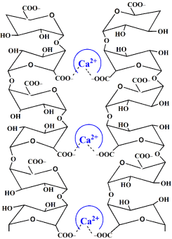
Figure 1.
Schematic representation of two homogalacturonan chains forming complex with calcium ions.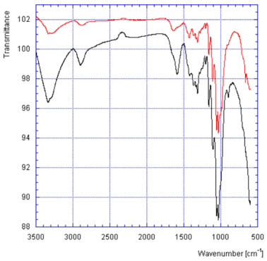
Figure 2.
ATR FTIR spectra of the Shroud body image fiber from the 1EB sticky type (black, lower plot) and a non-image fiber from the Shroud corner (red, upper plot). Reproduced from G. Fanti paper [37] with written permission from the author.